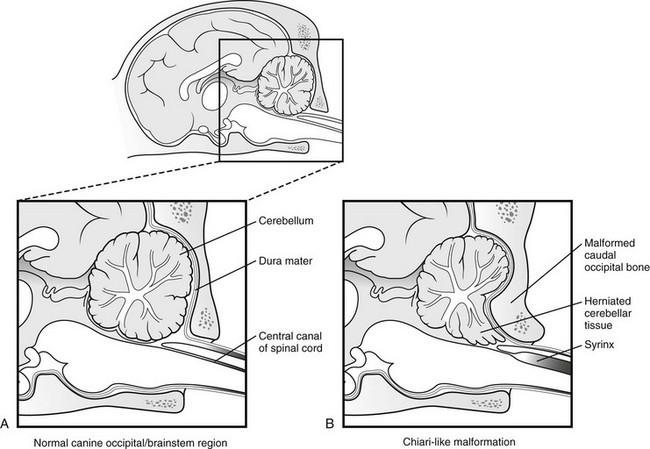
Caudal Occipital Malformation Syndrome (COMS) is a congenital neurological disorder predominantly observed in certain dog breeds, particularly the Cavalier King Charles Spaniel. This syndrome is characterized by malformations in the caudal occipital region, where the skull meets the spine, leading to a range of neurological and structural abnormalities.
Clinical manifestations of COMS in dogs commonly include cerebellar herniation through the foramen magnum, which is the opening at the base of the skull. This herniation can result in the formation of syringomyelia, fluid-filled cavities within the spinal cord. Additionally, dogs affected by COMS may exhibit abnormalities in the shape of the skull.
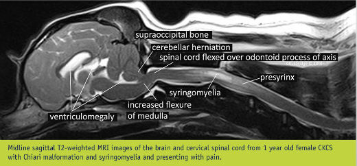
Clinical signs and symptoms vary but often include pain, ataxia (lack of coordination), difficulty swallowing, and weakness. Diagnosis typically involves advanced imaging techniques such as magnetic resonance imaging (MRI) to visualize the structural anomalies in the caudal occipital region and assess the severity of cerebellar herniation.
Treatment options for COMS depend on the severity of the condition. Medical management may be employed to alleviate pain, while surgical interventions such as craniotomy or foramen magnum decompression aim to relieve the compression on the spinal cord and correct the malformations. The success of treatment and prognosis are influenced by the extent of neurological damage and the effectiveness of the chosen interventions.
Managing COMS in dogs requires a collaborative effort involving veterinary neurologists, surgeons, and rehabilitation specialists. Long-term care and monitoring are often necessary to address persistent neurological symptoms and ensure the overall well-being of affected dogs. Early detection and intervention play a crucial role in optimizing outcomes for dogs diagnosed with Caudal Occipital Malformation Syndrome.
The diagnosis is made by visualizing the defect with MRI and also ruling-out other common diseases that can cause the same clinical signs. Because this defect is commonly identified and is not always the cause of significant problems, a spinal tap is often performed to address the possibility of another common disease in small breed dogs called meningoencephalitis.
Medical management of this condition is often with pain modulators like gabapentin (Neurontin) or pregabalin (Lyrica) for neuropathic pain, and other pain medication (opiates, NSAIDS or steroids) and medication to try to decrease fluid production (steroid, omeprazole (Prilosec), furosemide (Lasix)).
Surgery for this condition is called a foramen magnum decompression (FMD) and has the goal of alleviating the pinching of the spinal cord, smoothing out the flow of spinal fluid, and therefore eliminating the impetus for neuropathic pain and weakness. A FMD has a success rate of about 80% to improve the pain, weakness and vertigo. Many variables exist to explain why some dogs do better than others. Long term, there is a reported recurrence rate as high as 25% following surgery.
